Collagenase digestion method - culturing umbilical cord mesenchymal stem cells
Human umbilical cord mesenchymal stem cells have high differentiation potential, stable genes, are not susceptible to mutation, and do not cause tumors; they do not require matching and no rejection, and are widely used in disease treatment and anti-aging research. At present, the application in the beauty industry and for the sub-health groups is also attracting more and more attention.
The umbilical cord of umbilical cord mesenchymal stem cells is composed of capsule, Huatong gum, umbilical vein and umbilical artery umbilicus. Umbilical cord mesenchymal stem cells are present in Huatong's gum.
Due to the small amount, the amount of cells obtained after digestion with collagenase is small. In addition, when the collagen is obtained by the general collagenase digestion method, the collagenase is more harmful to the cells, and the cultured cells are in a poor state.
The patch method is a commonly used method for culturing human umbilical cord mesenchymal cells, but the cells climb out of the tissue for a long time (10-20 days or even longer). After continuous exploration, we found a way to obtain umbilical cord mesenchymal stem cells by collagenase digestion.
The specific operation method is as follows:
1 After cutting the baby's umbilical cord and the mother's umbilical cord, immediately clamp it with the umbilical cord clamp to prevent internal contamination. Load into a sterile bag (container) and transfer to the laboratory.
2 In the biosafety cabinet or in the clean bench, remove the umbilical cord and use a 75% alcohol soak for 2 minutes (50ml centrifuge tube or 250px dish). If the umbilical cord is long and cannot be loaded into the container, the umbilical cord can be cut to about 250px, but the umbilical cord clamp must be added before cutting to prevent alcohol from entering the umbilical cord and damaging the cells.
3 After treatment with alcohol, quickly transfer it to a container filled with PBS or saline for 2 times, 2 minutes each time.
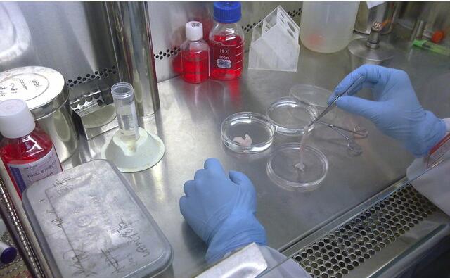
4 Cut off the umbilical cord clip, cut the umbilical cord into 2-75px segments, use the scissors to cut the umbilical cord from the inside of the umbilical vein, and use the scorpion scorpion (the umbilical cord tissue is relatively slippery, with the scorpion scorpion to operate) to the umbilical vein, umbilical artery and umbilical cord. The envelope is removed to prevent the incorporation of miscellaneous cells.
5 Cut the umbilical cord tissue with scissors, dilute 0.1% type II collagenase (dilution solution with DMEM/F12) overnight, and digest overnight at 4 °C.
6 The next day, pour the undigested tissue onto a 100 mesh (80 mesh, 160 mesh) steel screen (preferably a steel mesh screen, carrying a large amount of tissue for easy operation), with PBS (or saline) rinse multiple times to remove as much collagenase as possible from the surface of the tissue. Finally, rinse with 1-2 times of complete medium to remove PBS (or saline).
7 Pour into the dish (inconvenient to put in the bottle), add a small amount of medium culture on the undigested tissue block (6ml plus 2-3ml, 10ml plus 3-4ml) (the purpose is to reduce the residual collagenase on the cells) hurt). The next day, the tissue block completely disappeared (the collagenase immersed in the tissue was digested), the cells were completely dispersed, and under the microscope, there were many dispersed cells in the already digested tissue. A small amount of medium (150 px plus 2-3 ml, 250 px plus 4-5 ml) was added to the medium and the culture was continued.
8 After two days, continue to add a small amount of medium (150px plus 2-3ml, 250px plus 4-5ml).
9 After one or two days, the number of cells will increase significantly (many colony growth), transferred into two 50ml centrifuge tubes, add PBS (or saline) to 50ml, mix, centrifuge at 2500rpm, 6min.
10 Resuspend in 5-6 ml (depending on the amount of cells) in complete medium, centrifuge at 1000 rpm for 6 min, place in a T25 flask, and add 5 ml of complete medium.
After 13 days, the culture can be continued by changing the medium or adding 3-4 ml of complete medium. After every three days, change the liquid until it is over (more than 70% can be passed down)
Revelation notes:
1 This method is easy to operate and has a short incubation time.
2 Digestion of the umbilical cord with collagenase, at 37 ° C, the umbilical cord can be basically digested within a few hours, but at the same time the damage to the cells is greater. Collagenase can penetrate into the umbilical cord tissue, and low concentrations of collagenase digest tissue from the inside to the outside, causing less damage to cells. (Trypsin has very poor digestion effect on umbilical cord tissue)
3 The reason for completely digesting the tissue and not separating the cells separately is that the umbilical collagen component (viscous) promotes the proliferation of umbilical cord mesenchymal stem cells. Most of the cells are suspended in collagen and proliferate in colonies, which is the main reason why the cells are kept in a good condition.
4 When culturing umbilical cord mesenchymal stem cells, do not use serum-containing medium (serum promotes cell differentiation), but use stem cell-specific medium. Passage is also try not to trypsin digestion (trypsin damage to umbilical cord mesenchymal stem cells), special digestive juice should be used to ensure more passage times.
Drape Pack,Vertical Drape Pack,Disposable Drape Pack,Disposable Medical Drape Pack
ShaoXing SurgeCare Medical Products Co., Ltd. , https://www.sxsurgecaremedical.com